
|
| OVERVIEW |
| Flythrough Tour |
| ~~~~~~~~~~~~ |
| MOLECULAR PROCESSES |
| Transcription |
| Regulated Transcription |
| mRNA Processing |
| mRNA Splicing |
| Translation |
| Lac Operon |
| ~~~~~~~~~~~~ |
| CELLULAR PROCESSES |
| Protein Trafficking |
| Protein Modification |
| Protein Recycling |
| Insulin Signaling |
| Constitutive Secretion |
| Regulated Secretion |
| Mitochondrial Protein Transport |
| Mitosis |
| Meiosis |
| ~~~~~~~~~~~~ |
| CELLULAR ENERGY CONVERSION |
| Atp Synthase (Gradients) |
| Electron Transport Chain |
| Photosynthesis (Light Reaction) |
| Photosystem II |
| Glycolysis (Overview) |
| Glycolysis (Reactions) |
| Citric Acid Cycle (Overview) |
| Citric Acid Cycle (Reactions) |
| Energy Consumption |
| ~~~~~~~~~~~~ |
|
| ~~~~~~~~~~~~~ |
| NDSU Virtual Cell YouTube |
| MCBE Home |
| Virtual Cell |
| WWWIC Home |
| Funding & Credits |
| ~~~~~~~~~~~~~ |


| HOME > INSULIN SIGNALING > ADVANCED LOOK > 1.) CELL MEMBRANE |
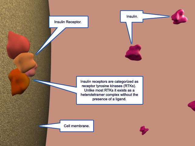
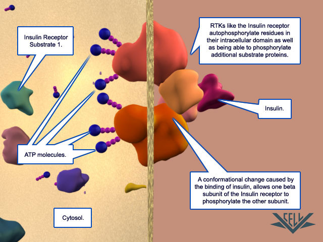
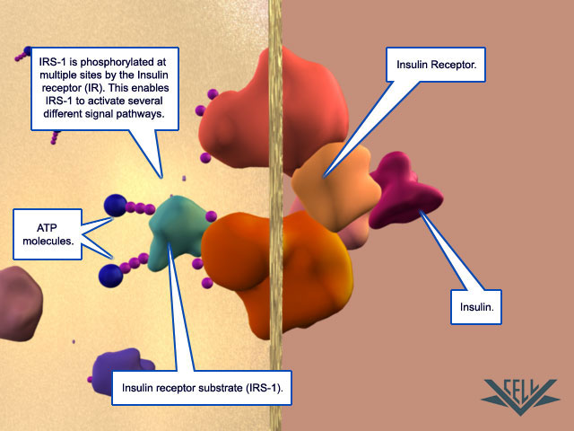
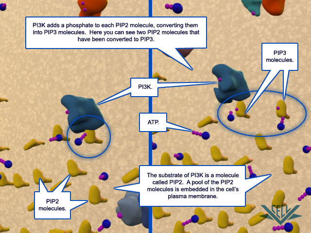
| Insulin Signaling:
Advanced Look -->
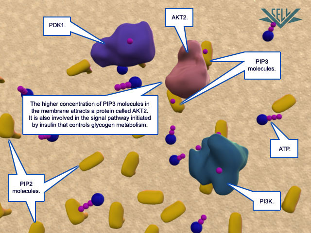
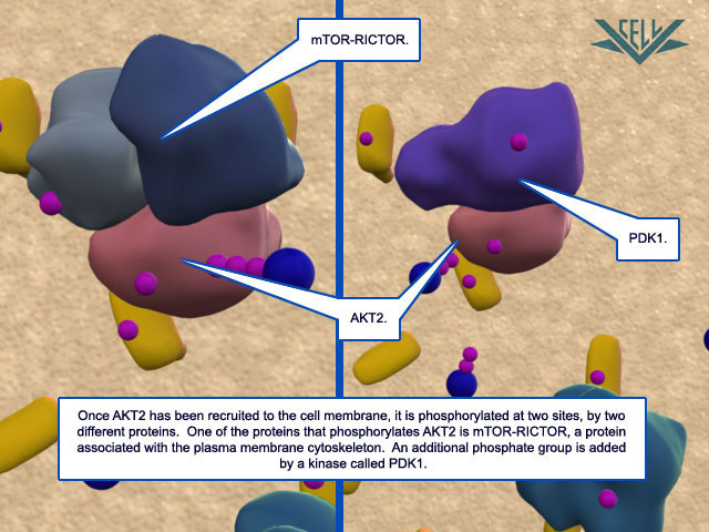
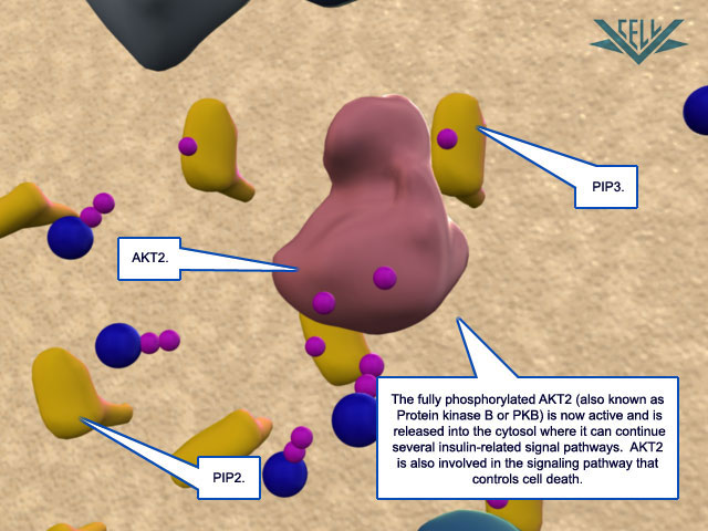
1.) Cell Membrane
Most of the phosphorylation signals involved in this insulin signal pathway occur at the cell membrane. Clicking on each of the thumbnail images will bring up a larger, labeled version of the described scene. To see the Flash movie for the following sequence of images, click here.
NEXT --> 2.) GSV POOL |


This work is licensed under a Creative Commons Attribution-NonCommercial-NoDerivatives 4.0 International License.